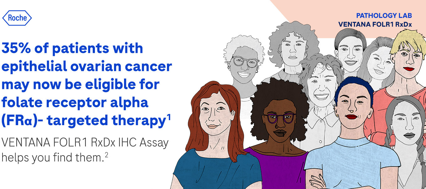
35% of patients with epithelial ovarian cancer may now be eligible for folate receptor alpha (FRα)- targeted therapy1
VENTANA FOLR1 RxDx IHC Assay helps you find them.2
VENTANA FOLR1 RxDx IHC Assay helps you find them.2

Introducing the VENTANA FOLR1 (FOLR1-2.1) RxDx Assay, the first FDA-approved immunohistochemistry companion diagnostic test that helps you detect the Folate Receptor (FOLR1) biomarker in patients with epithelial ovarian cancer (EOC).2 By testing for FR alpha expression, you can help identify 35% of patients with EOC that may benefit from a promising FR alpha-targeted therapy.1
As part of its ongoing commitment to cancer care, Roche is proud to offer this advanced diagnostic tool, giving labs a way to help meet a critical need of patients with EOC.
Intended Use2
VENTANA FOLR1 (FOLR1-2.1) RxDx Assay is a qualitative immunohistochemical assay using mouse monoclonal anti-FOLR1, clone FOLR1-2.1, intended for use in the assessment of
folate receptor alpha (FOLR1) protein in formalin-fixed, paraffin-embedded epithelial ovarian, fallopian tube, or primary peritoneal cancer tissue specimens by light microscopy.
This assay is for use with OptiView DAB IHC Detection Kit for staining on a BenchMark ULTRA instrument.
FOLR1 expression clinical cut-off is ≥ 75% viable tumor cells (TC) with membrane staining at moderate and/or strong intensity levels.
This assay is indicated as an aid in identifying patients with epithelial ovarian, fallopian tube, or primary peritoneal cancer who may be eligible for
treatment with ELAHERE (mirvetuximab soravtansine).
Test results of the VENTANA FOLR1 (FOLR1-2.1) RxDx Assay should be interpreted by a qualified pathologist in conjunction with histological examination,
relevant clinical information, and proper controls.
This product is intended for in vitro diagnostic (IVD) use.
Exhibits 98% moderate and strong membrane staining or ≥ 75% tumor cells membrane staining with complete circumferential pattern

Caris Life Sciences
4610 South 44th Place,
Phoenix, AZ 85040
Phone: 602-464-7500

NeoGenomics Laboratories
31 Columbia Aliso Viejo, CA 92656
Phone: 866-776-5907, option 3

LMC Pathology Services
7455 W. Washington Avenue,
Suite 301, Las Vegas, NV 89128
Phone: 877-LMC-LABS
Phone: 702-732-3441
Fax: 702-732-2310
Labcorp
3 Forest Parkway
Shelton, CT 06484
Phone: 800-345-GENE (4363)
Fax: 888-287-8199
The VENTANA FOLR1 (FOLR1-2.1) RxDx Assay Interpretation Guide offers valuable information to pathologists in the appropriate use of this product.
Your guide provides:
Scoring information
Staining characteristics
Additional guidance
*Images of primary peritoneal and primary fallopian tube cancer are not included in visual illustrations due to the rarity of these indications.